|
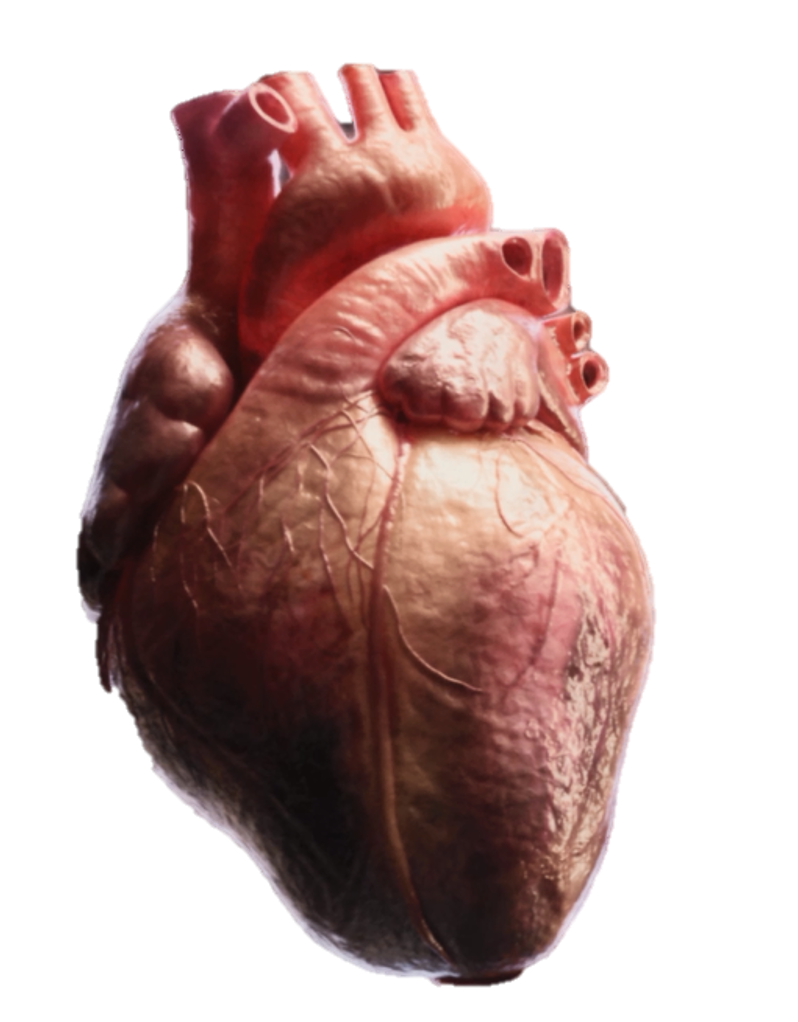
About our research.The mammalian heart is composed of a number of cell types including muscle cells and fibroblasts. Cardiac muscle cells are interconnected at their ends through their intercalated discs and are composed of intercellular junctions (gap junctions, desmosomes and area composita) essential for maintaining correct contraction of the heart. Cardiac fibroblasts produce and degrade extracellular matrix (ECM) components. The ECM acts as the structural network and signaling mediator in the heart.
|
We focus on the in vitro engineering of functional
myocardium that mimics human heart tissue for analysis of
myocardial function
- We differentiate human pluripotent stem cells into functional cardiomyocytes and study different genetic heart diseases (for instance Marfan syndrome) with the help of induced pluripotent stem cell (iPSC) technology.
- Dysfunction may result from a complicated interaction of various cell types, thus more complex heart models are needed. Our objective is to mimic this interaction by realizing 3D co-culture of all cell types that make up the heart, including the fibroblasts that synthesize the ECM, to get a deeper understanding of this interaction.
- We conduct research with biomaterials suitable for the cultivation of cardiomyocytes and develop different ways to characterize the functional properties of the cardiomyocytes.
We integrate these in vitro heart models with (genetically
modified) mouse models
- Cells communicate with each other and with the ECM. The cell’s internal cytoskeleton is physically connected via protein-protein interactions to other cells and the ECM. We have studied responsible interactor proteins (for instance alpha-catenins, beta-actin, cadherins).
- We investigate the functionality, morphology, histology, and ultrastructural features of interactor protein-null murine hearts, also we use cardiomyocytes derived from interactor protein-null stem cells.
- The aim of our work is not only to develop 3D in vitro cultures but also make 3D reconstructions of intercellular junction of the intercalated discs obtained from mouse models. We demonstrated that volume scanning electron microscopy revealed the close relation between gap junctions and desmosomes and their spatial distribution in a 3D manner.
We co-operate with experts in various fields, including engineers, experts in biomaterials, electrophysiologists, geneticists and clinicians.
CUSTOMEAT.From in vitro culturing of stem cells towards meat design.
The CUSTOMEAT project is a collaborative, fundamental research initiative (type SBO) aimed at expanding knowledge on research gaps and providing the technologies necessary to produce structured in vitro meat. This is achieved by integrating expertise from biomedical engineering with food technology know-how. Various types of stem cells, including embryonic stem cells and myoblast stem cells, are being investigated for cultured meat production. Additionally, efforts are focused on creating a food-grade matrix to which cells can adhere and that is capable of absorbing healthy nutrients contributing to the flavor, texture, and color of the meat. The produced muscle cells and matrix are then combined via tissue engineering to create muscle tissue. Beyond the technical aspects of production, the project also examines the societal impact of cultured meat, including consumer acceptance and perception, environmental impact, and regulatory considerations.
Click this link to learn more about this project. |


|
We focus on the in vitro engineering of functional bovine muscle tissue to advance the production of structured cultured meat
- We produce embryonic stem cell lines via IVF and characterizing them for pluripotency, trilineage differentiation potential, and genomic stability.
- We develop bovine myoblast cell lines characterized by CD56 to ensure their myogenic potential and suitability for muscle tissue engineering.
- We differentiate embryonic stem cells and myoblast stem cells into muscle cells, aiming to enhance our understanding of myogenesis and improve muscle tissue maturation for cultured meat production.
- We study muscle tissue differentiation and maturation within a 3D environment, aiming to create a more complex environment and apply stimulation methods to improve myogenesis.
- We explore and test different food-grade biomaterials that support cell adhesion and nutrient absorption, which are crucial for developing muscle tissue with desirable flavor, texture, and color.
- Our research also includes the characterization of the mechanical and biochemical properties of the engineered muscle tissues, ensuring they meet the standards required for cultured meat production.
- We cooperate with experts in various fields, including tissue engineering groups, reproduction units, engineers, genetics and companies in the biomedical equipment and food sectors, to leverage their expertise and technology.