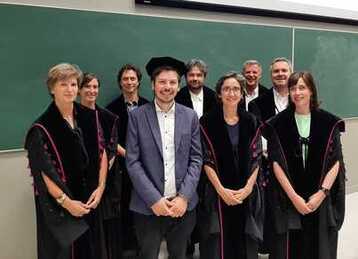
 Laurens Léger successfully defended his PhD thesis, titled 'One cell type, infinite possibilities: utilizing human pluripotent stem cells for modelling arrhythmogenic cardiomyopathy and cardiac tissue engineering', and obtained the degree of Doctor in Health Sciences. His PhD was supervised by prof. Jolanda van Hengel, together with Dr. Martina Calore.
Laurens was a valued member of the team, his input helped broaden the expertise and knowledge of the Medical Cell Biology group. We wish him the best of luck on his future endeavours!
0 Comments
 Our lab succesfully generated an iPSC line from a patient suffering from Marfan syndrome (carrying a heterozygous c.7754T>C variant in FBN1). Additionally, an isogenic control was generated using CRISPR/Cas9 technology. These iPSCs can be readily differentiated into cell types of interest, such as vascular smooth muscle cells, and cardiomyocytes.
This isogenic pair will help advance fundamental knowledge concerning Marfan Syndrome. You can access the paper using this link. The first CorEuStem (European COST action surrounding stem cells) short term scientific mission allowed hands-on training on hPSC culture, primary cells transduction towards pluripotency and small molecule screening using hPSCs. Karina Goluba, research assistant from University of Latvia, Riga (Latvia) was introduced to the Medical Cell Biology lab run by Professor Jolanda van Hengel, where she learned how to reprogram somatic cells into iPSCs, basic procedures regarding hPSCs maintenance and differentiation.  “Overall, during this mission I learned all the planned methods and fully implemented the goals set at the beginning. I have gained very good experience in working with pluripotent stem cells…and now I can feel confident working with them soon in my PhD work and in other projects. This STMS also helped establish scientific connection between our host institutions.” Good news! Our paper named Microscopic visualization of Cell-Cell Adhesion Complexes at Micro and Nanoscale was published in Fronties in Cell and Developmental Biology, April 20th 2022.  In this review, we discuss the main light and electron microscopy techniques used to unravel the structure and composition of the three cell-cell contacts in epithelial and endothelial cells. It functions as a guide to pick the appropriate imaging technique(s) for the adhesion complexes of interest. We also point out the latest techniques that have emerged. At the end, we discuss the problems investigators encounter during their cell-cell adhesion research using microscopic techniques.
Read the paper by clicking this link.  We are proud to announce that our co-authored manuscript titled photothermal nanofibres enable safe engineering of therapeutic cells was published on October 21st in Nature Nanotechnology.
This paper showed that cell membrane permeabilization with photothermal nanofibres is a promising concept towards the safe and more efficient production of engineered cells for therapeutic applications, including stem cell or adoptive T cell therapy. Access the full article by clicking this link.  Our review titled 'Time for rethinking the different Beta actin transgenic mouse models?' has been accepted for publication in the journal 'cytoskeleton'.
The article can be accessed by clicking here. A short abstract can be found below: The actin family is crucial for many cellular processes and in mammals muscle and non‐muscle forms exist. The latter group contains cytoplasmic‐β‐actin and cytoplasmic‐γ‐actin, almost identical in amino acid sequence and with a significant functional overlap. We introduce the properties of the Actb gene and mRNA transcript(s) with main focus on the 3′UTR and its unique features, that is, the zipcode and two polyadenylation sites creating transcripts of different lengths. Several transgenic mouse models with a modified Actb locus have been created. Whole body knockouts and, with one exception, insertion models lead to embryonic lethality indicating that the Actb gene or transcripts or translated β‐actin are essential. Tissue specific ablation at later developmental stages lead to no, or mild phenotypes, suggesting that the Actb gene or β‐actin protein is somewhat dispensable. Gene edited Actb mice that produce γ‐actin are viable. This assumes that the nucleotide sequence of Actb is important and not the specific amino acid sequence of the protein it encodes. Upregulation of other actin paralogs was frequently observed upon β‐actin ablation and can also engage in the phenotype. For a better understanding, it will be necessary to analyze in current and future models all relevant actin transcripts and protein levels in a standardized and comprehensive way. |
 RSS Feed
|
Medical Cell Biology lab
|