 On the 7th of October, our article titled: "Effects of fibrillin mutations on the behavior of heart muscle cells in Marfan syndrome" was published in the journal Scientific Reports, Nature.
The article describes the first in vitro cardiomyocyte model of Marfan Syndrome. You can access the article for free by clicking here. A short abstract can be read below: Marfan syndrome is a systemic disorder caused by defects in fibrillin-1, a matrix protein. Cardiovascular manifestations, particularly in the aorta, are the most life-threatening consequences. Accumulating evidence from patients and mouse models indicates that Marfan syndrome is also causing a primary cardiomyopathy, but little is known about the mechanism. In this study, induced pluripotent stem cells derived from a Marfan syndrome patient were differentiated to heart muscle cells. This provides a unique alternative approach to study Marfan cardiomyopathy. Here we report the first and only cardiac cell culture model for Marfan syndrome, revealing abnormalities in the behavior of Marfan heart muscle cells that are related to matrix defects. Based on these results, we postulate that impaired support from the extracellular environment plays a key role in the improper functioning of heart muscle cells in Marfan syndrome.
0 Comments
We published the following paper: "Liquid marble technology to create cost-effective 3D cardiospheres as a platform for in vitro drug testing and disease modelling." in the journal MethodsX. Three-dimensional (3D) cell culturing has several advantages over 2D cultures. 3D cell cultures more accurately mimic the in vivo environment, which is vital to obtain reliable results in disease modelling and toxicity testing. Current methods to achieve multicellular spheroids are expensive, time-consuming and require specialized materials and training. Hydrophobic powders can be used to create a micro environment for cell cultures, which are termed liquid marbles (LM). In this procedure we describe the first use of the LM technology for 3D culturing in vitro derived human cardiomyocytes which results in the formation of cardiospheres within 24h. The cardiospheres could be used for several in depth and high-throughput analysis. You can access the article by clicking here. 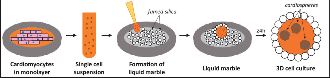 Bieke Vanslembrouck succesfully obtained her PhD degree on the 7th of May. The title of her dissertation was: Exploring the three dimensional organisation of the murine intercalated disc and unravelling the connection between the intercalated discs and the cytoskeleton of cardiac myocytes. We wish her the best of luck on her future endeavours in the group of Carolyn Larabell, Lawrence Berkeley National Laboratory.  Robust protocol for feeder-free adaptation of cryopreserved human pluripotent stem cellsOn the 29th of october, a new paper was published in 'In Vitro Cellular & Developmental Biology - Animal'. For a link to the article, click here. (note: this is now open access!)
You can find the abstract below. Human pluripotent stem cells (hPSCs) are conventionally maintained on mouse embryonic fibroblast (MEF) feeder layers. However, downstream applications, such as directed differentiation protocols, are primarily optimized for feeder-free cultures. Therefore, hPSCs must often be adapted to feeder-free conditions. Here we propose a novel feeder-free adaptation protocol using StemFlex medium, which can be directly applied to thawed hPSC lines. The direct feeder-free adaptation protocol using StemFlex culture medium on Geltrex coating led to robust hPSC cultures in approximately 2 weeks. This approach was tested with three human embryonic stem cell (hESC) lines. All lines were confirmed to be pluripotent, expressing POU5F1, SOX2, and NANOG. No chromosomal imbalances were induced by the feeder-free adaptation. StemFlex medium enabled the efficient adaptation of hPSCs to feeder-free conditions directly after thawing. This protocol is easy to implement in laboratories that perform feeder-free cultures, allowing more convenient adaptation and more robust expansion of cryopreserved hPSCs, even in cases when sample quality is low or unknown.  On sunday, the 24st of november, the annual Day of Science (Dag van de Wetenschap) is taking place. Our laboratory also hosts a guided tour, where young and old can learn about cardiovascular research. Additional info about the event can be found on the official webpage.
|
 RSS Feed
|
Medical Cell Biology lab
|